Specialized Neuro-Ophthalmology Care in Kharghar, Navi Mumbai
Neuro-ophthalmology services in Kharghar, Navi Mumbai at Utsav Eye Clinic provides specialized diagnosis and management for vision disorders linked to the nervous system. Under double-fellowship trained specialist Dr. Anand Kumar, we treat double vision (diplopia), optic neuritis, papilledema (Pseudotumor Cerebri), ptosis, nystagmus, and unexplained vision loss. Call 022 35569270.
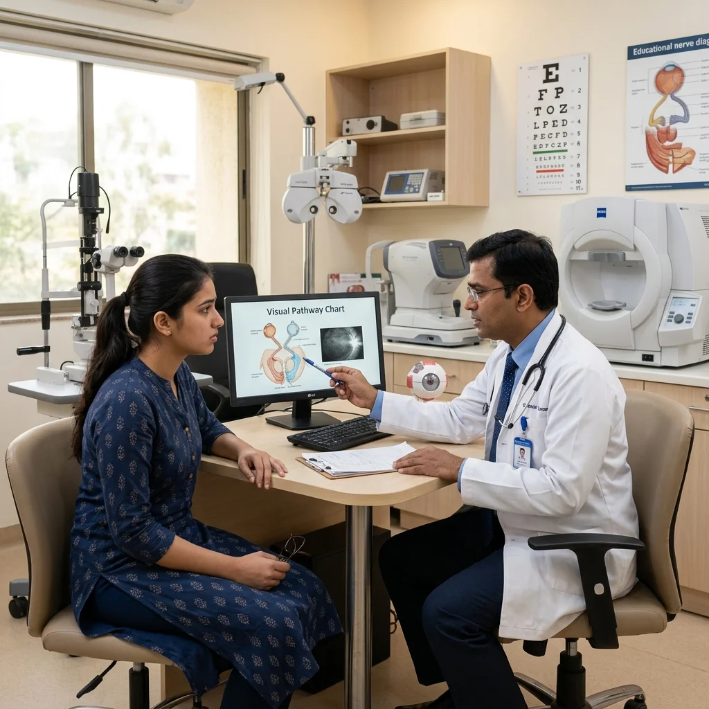
Many visual problems do not originate in the eye structure itself, but rather in the complex electrical connections between the eye and the brain. The optic nerve acts as a major cable carrying over a million individual visual fibers. Any inflammation, compression, stroke, or injury along this pathway can cause devastating visual loss or double vision. Neuro-ophthalmology is the medical subspecialty dedicated to diagnosing and managing these complex conditions.
What is Neuro-Ophthalmology?
The human visual system is incredibly complex. The eyes capture light, but it is the brain that actually "sees." The neural visual pathway extends from the retina at the back of the eye, runs through the long optic nerves, crosses at the optic chiasm, and travels to the visual cortex in the occipital lobe at the very back of the brain.
Neuro-ophthalmologists have super-specialized training to evaluate visual loss from a neurological perspective. They assess how the optic nerve, pupillary reflexes, and the six muscles controling eye movements interact with the central nervous system. Because many neuro-ophthalmic symptoms can be warning signs of serious systemic or neurological disorders, accurate diagnosis is critical.
Conditions We Diagnose and Manage
At Utsav Eye Clinic, we treat a comprehensive spectrum of neuro-ophthalmic conditions in adult and pediatric patients:
- Optic Neuritis: Inflammation of the optic nerve that causes sudden, painful visual dimness and reduced color vision, often representing the first sign of demyelinating diseases like Multiple Sclerosis (MS) or Neuromyelitis Optica (NMO).
- Papilledema & Raised Intracranial Pressure: Swelling of the optic nerve discs in both eyes due to elevated pressure inside the skull, which can result from brain swelling, tumors, or **Idiopathic Intracranial Hypertension (Pseudotumor Cerebri)**.
- Double Vision (Diplopia): Seeing two of a single object, typically caused by misalignment of the eyes. This can result from cranial nerve palsies (3rd, 4th, or 6th nerve palsies) affecting the eye muscles.
- Ischemic Optic Neuropathy: A "stroke" of the optic nerve, caused by a sudden lack of blood supply, leading to acute, painless visual loss (e.g., Anterior Ischemic Optic Neuropathy - AION).
- Optic Nerve Compression: Loss of vision or visual fields (such as bitemporal hemianopia) caused by pituitary adenomas, meningiomas, or other skull-base tumors compressing the optic chiasm.
- Myasthenia Gravis: An autoimmune neuromuscular junction disorder that causes fluctuating weakness in muscles, frequently presenting as drooping eyelids (ptosis) and variable double vision.
- Blepharospasm & Hemifacial Spasm: Involuntary, painful eyelid twitching or squeezing that can severely impact a patient's ability to keep their eyes open.
- Pupillary Abnormalities: Unexplained differences in pupil size (anisocoria), sluggish pupil reactions, or conditions like Adie's tonic pupil and Horner's syndrome.
- Unexplained Vision Loss: Visual deficits that cannot be accounted for by typical ocular issues like cataracts, refractive errors, or macular disease, requiring neural pathway mapping.
Neuro-Ophthalmology Diagnostic Techniques
Diagnosing a neuro-ophthalmic condition requires a meticulous, systematic clinical examination coupled with advanced diagnostic imaging. At Utsav Eye Clinic, our facility is equipped with:
-
1. Automated Visual Field Analyzer (HFA):
Essential for mapping your peripheral and central vision. Specific patterns of visual field loss (such as hemianopias or quadrantanopias) help pinpoint the exact location of lesions along the visual pathway in the brain.
-
2. High-Resolution Optical Coherence Tomography (OCT):
Provides cross-sectional, microscopic views of the optic nerve head and the retinal nerve fiber layer (RNFL). OCT allows us to measure thinning of the nerve fibers (optic atrophy) or swelling (papilledema) with micron-level precision.
-
3. Digital Fundus Photography:
Captures high-resolution color photographs of the optic nerve disc to document and monitor changes in swelling, pallor, or cupping over time.
Why Specialized Neuro-Ophthalmology Care Matters
Unlike standard eye conditions that can be treated with glasses or routine surgeries, neuro-ophthalmic disorders often involve systemic, vascular, or neurological conditions. A specialized approach is critical because:
- Early Tumor Detection: Compressed visual fields or unexplained vision loss can be the very first sign of a pituitary tumor or brain aneurysm. Prompt diagnosis can be life-saving.
- Preventing Optic Atrophy: Swollen or inflamed optic nerves will eventually progress to permanent nerve fiber loss (atrophy) if pressure is not reduced or inflammation is not controlled.
- Collaborative Treatment: We work in close coordination with leading neurosurgeons, neurologists, endocrinologists, and radiologists in Navi Mumbai to provide comprehensive care.
Frequently Asked Questions (FAQ)
What is the difference between an ophthalmologist and a neuro-ophthalmologist?
A general ophthalmologist deals with structural eye issues like cataracts, glaucoma, and dry eyes. A neuro-ophthalmologist has super-specialty fellowship training to diagnose and treat visual problems that originate in the brain, nervous system, and cranial nerves.
Can double vision be cured without surgery?
Yes, many cases of double vision (diplopia) can be managed non-surgically using temporary prism lenses on glasses, specialized eye exercises, patching, or treating the underlying medical condition (like high blood pressure or diabetes). If it persists, eye muscle realignment surgery is performed.
What is optic neuritis and is it curable?
Optic neuritis is inflammation of the optic nerve, often causing sudden blurred vision and pain with eye movement. It is highly treatable, often resolving on its own or accelerated with intravenous steroid therapy, with most patients recovering baseline vision.
What causes papilledema?
Papilledema is swelling of both optic nerves caused by increased pressure inside the skull (intracranial pressure). Causes include brain tumors, meningitis, hydrocephalus, or Idiopathic Intracranial Hypertension (Pseudotumor Cerebri). It requires immediate investigation.
What is Pseudotumor Cerebri?
Also known as Idiopathic Intracranial Hypertension (IIH), this condition mimics the symptoms of a brain tumor (headaches, double vision, optic nerve swelling) but without a physical tumor. It is treated with medications, weight management, and careful visual monitoring.
Why do I need an MRI scan for an eye problem?
An MRI scan is ordered when a visual problem originates along the optic nerve pathway or in the brain's visual cortex. It helps rule out nerve inflammation, compression from tumors, or vascular issues that standard eye exams cannot see.
Can a stroke cause vision loss?
Yes. A stroke affecting the visual pathways in the brain can cause sudden loss of part of your visual field (e.g., hemianopia), double vision, or eye movement control issues. Neuro-ophthalmologists assess and help rehabilitate these visual deficits.
What is Myasthenia Gravis and how does it affect the eyes?
Myasthenia Gravis is an autoimmune disorder that causes muscle weakness. In the eyes, it commonly causes fluctuating drooping of the upper eyelids (ptosis) and double vision, which worsen as the day progresses or with fatigue.

MBBS, MS (Ophthalmology), Fellowship in Pediatric Ophthalmology & Strabismus (USA). Chief Surgeon & Director at Utsav Eye Clinic.
Utsav Eye Clinic in Kharghar Sector 21 is easily accessible to patients across Navi Mumbai, including Belapur, Panvel, Vashi, Nerul, and Kamothe.