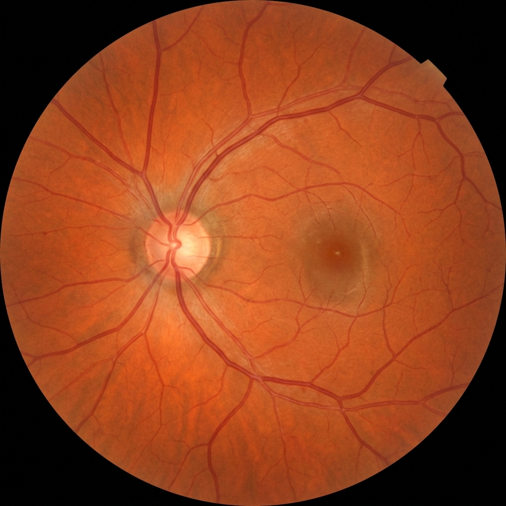
What Is the Retina?
The retina is a thin layer of light-sensitive tissue lining the back of the eye. It functions like the film in a camera — it captures light rays focused by the cornea and lens, converts them into neural signals, and sends them to the brain via the optic nerve. The central part of the retina, called the macula, is responsible for sharp, detailed central vision used for reading, recognising faces, and driving.
Because the retina is vital for vision, any damage or disease affecting it can lead to significant visual impairment or even blindness if not detected and treated early. Regular comprehensive eye examinations — especially for adults over 40, diabetics, and those with a family history — are essential for protecting your retinal health.
Common Retina Disorders
1. Diabetic Retinopathy
Diabetic retinopathy is the most common cause of vision loss among people with diabetes and a leading cause of blindness in working-age adults. Chronically high blood sugar levels damage the tiny blood vessels in the retina, causing them to leak fluid, bleed, or close off entirely.
The condition progresses through two stages:
-
🟡
Non-Proliferative Diabetic Retinopathy (NPDR):
The early stage. Tiny blood vessels (microaneurysms) weaken and leak, causing retinal swelling (macular oedema). Vision may be normal or mildly blurred at this stage.
-
🔴
Proliferative Diabetic Retinopathy (PDR):
The advanced stage. The retina grows new, abnormal blood vessels (neovascularisation) that are fragile and bleed into the vitreous. This can cause sudden vision loss, scar tissue, and retinal detachment.
Treatment: Depending on severity, treatment includes intravitreal anti-VEGF injections (bevacizumab, ranibizumab, aflibercept) to reduce swelling and abnormal vessel growth, laser photocoagulation to seal leaking vessels, and vitrectomy surgery for advanced cases with vitreous haemorrhage.
2. Age-Related Macular Degeneration (AMD)
AMD is the leading cause of irreversible vision loss in people over 60. It affects the macula, causing loss of sharp central vision. There are two forms:
-
👁
Dry AMD (Atrophic):
The more common form (80–90% of cases). Yellow deposits called drusen accumulate under the retina, and the macular tissue gradually thins and atrophies. Vision loss is usually gradual. There is no cure, but AREDS2 nutritional supplements (vitamins C, E, zinc, lutein, zeaxanthin) can slow progression in intermediate cases.
-
👁
Wet AMD (Neovascular):
Less common but more severe. Abnormal blood vessels grow under the retina and leak blood or fluid, rapidly distorting or destroying central vision. Treated with anti-VEGF injections, which can stabilise or even improve vision when started early. Self-monitoring with an Amsler grid helps detect early changes.
3. Retinal Detachment
Retinal detachment is an ophthalmic emergency where the retina peels away from its underlying support tissue. Without prompt treatment, it can lead to permanent vision loss in the affected eye. Risk factors include high myopia, prior eye surgery, trauma, and a family history of detachment.
Symptoms to watch for: Sudden increase in floaters (dark spots or lines), flashes of light (photopsia), and a shadow or curtain descending over part of the visual field. If you experience any of these, seek immediate eye care.
Treatment: Surgical options include pneumatic retinopexy (gas bubble injection), scleral buckle (silicone band around the eye), and vitrectomy (removal of vitreous gel). The choice depends on the type, location, and severity of the detachment.
4. Retinal Vein Occlusion (RVO)
Retinal vein occlusion occurs when a blood clot blocks a vein in the retina. It is the second most common retinal vascular disease after diabetic retinopathy. There are two types:
- Branch Retinal Vein Occlusion (BRVO): A smaller branch vein is blocked, affecting part of the visual field.
- Central Retinal Vein Occlusion (CRVO): The main retinal vein is blocked, leading to more extensive visual loss.
Risk factors include high blood pressure, diabetes, glaucoma, and blood clotting disorders. Treatment typically involves anti-VEGF injections and, in some cases, laser therapy to reduce macular oedema.
5. Epiretinal Membrane & Macular Hole
An epiretinal membrane (also called macular pucker or cellophane maculopathy) is a thin sheet of scar tissue that forms on the surface of the macula. It can contract and wrinkle the retina, causing distorted or blurred central vision. A macular hole is a small break in the macula, often caused by vitreous traction. Both conditions are more common after age 50 and may require vitrectomy surgery if they significantly affect vision.
Warning Signs — When to See a Doctor Immediately
Seek urgent ophthalmic evaluation if you experience any of the following:
- ⚠️ Sudden increase in floaters or dark spots in your vision
- ⚠️ Flashes of light, especially in peripheral vision
- ⚠️ A shadow or dark curtain spreading across your visual field
- ⚠️ Sudden painless loss of vision in one eye
- ⚠️ Distorted or wavy lines when looking at straight edges (e.g., door frames)
- ⚠️ A blank or dark spot in the centre of your vision
Retina Care at Utsav Eye Clinic
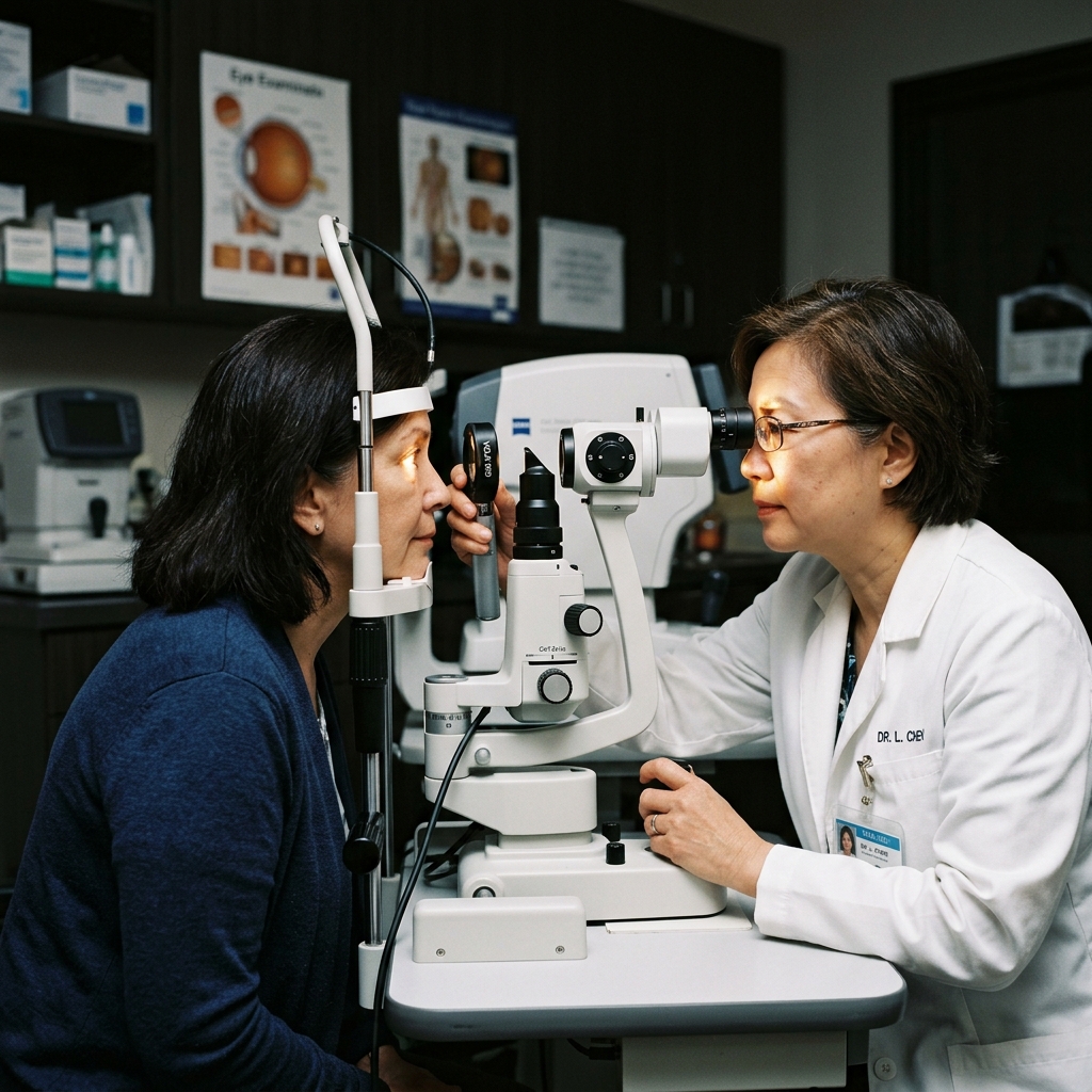
At Utsav Eye Clinic in Kharghar, Navi Mumbai, we provide comprehensive retina evaluation and management using advanced diagnostic tools including:
- Optical Coherence Tomography (OCT): High-resolution cross-sectional imaging of retinal layers to detect oedema, drusen, membranes, and holes.
- Digital Fundus Photography: Detailed colour photographs of the retina for documentation and monitoring of disease progression.
- Indirect Ophthalmoscopy: Comprehensive dilated examination of the entire retina, including the periphery.
Early detection is the cornerstone of preserving vision in retinal disease. If you are diabetic, over 50, highly myopic, or have a family history of retinal problems, schedule a comprehensive retinal screening today.
Frequently Asked Questions
Can diabetic retinopathy be reversed?
While existing damage cannot be fully reversed, early treatment with anti-VEGF injections and laser therapy can stop progression, reduce swelling, and in many cases improve vision. The most important step is to control blood sugar, blood pressure, and cholesterol levels.
Who is at risk for retinal detachment?
People with high myopia (nearsightedness above −6 dioptres), those who have had previous eye surgery (especially cataract surgery), individuals with a family history of retinal detachment, and people who have experienced eye trauma are at higher risk.
How often should I get my retina checked?
Adults over 40 should have a dilated eye exam every 1–2 years. Diabetics should have an annual retinal screening regardless of age. Those with known retinal conditions may need more frequent monitoring as advised by their ophthalmologist.
Are anti-VEGF injections painful?
The eye is numbed with anaesthetic drops before the injection, so most patients feel only mild pressure. The procedure takes just a few minutes and is performed as an outpatient procedure. Mild redness or irritation may occur for a day or two afterwards.